Anatomy Of Ribs Posterior - Han 200- 7B The skeleton at College at Oswego - StudyBlue : Beyond the lateral border of the first rib, the subclavian a.
byAdrienne Black•
0
Anatomy Of Ribs Posterior - Han 200- 7B The skeleton at College at Oswego - StudyBlue : Beyond the lateral border of the first rib, the subclavian a.. ✅ www.animatedanatomy.com/ ✅ ◄◄◄click to buy our anatomical software and lessons i also talk about sternum. The angles of the ribs form the most posterior extent of the thoracic cage. It is an atypical rib and is an important anatomical landmark and is one of the borders of the superior thoracic aperture. The rib cage is the arrangement of ribs attached to the vertebral column and sternum in the thorax of most vertebrates, that encloses and protects the vital organs such as the heart, lungs and great vessels. The nomenclature of the costal veins is the same as the arteries.
The rib cage is made up of the thoracic vertebrae, which we already covered, twelve pairs of ribs, each connected to a vertebra, the costal cartilage from the back, the ribs angle down slightly. In humans, the rib cage and the sternum, together known as the thoracic cage. Anatomy of the calf (posterior leg). Be sure to subscribe to the visible body blog for more anatomy awesomeness! Head, neck, tubercle, and body of a rib.
M1 Anatomy Study Guide (2014-15 Cullen) - Instructor ... from classconnection.s3.amazonaws.com The most superior rib is designated rib 1 and it articulates with the t1 thoracic vertebrae. The thoracic vertebrae lie in the posterior wall of the thorax with twelve pairs of ribs attached to them. Is immediately posterior to (and closely associated with) the phrenic n. The number is the same in both males and females. The serratus posterior inferior draws the lower ribs backward and downward to assist in rotation and extension of the trunk. They exhibit costal facets on each side at the junction of the vertebral body and the pedicle and on the transverse processes. ✅ www.animatedanatomy.com/ ✅ ◄◄◄click to buy our anatomical software and lessons i also talk about sternum. Ribs eight to ten are the false ribs and are connected to the sternum indirectly via the cartilage of the rib above serratus posterior.
Each pair articulates with a different thoracic vertebra on the posterior side of the body.
The most superior rib is designated rib 1 and it articulates with the t1 thoracic vertebrae. Transitions into the axillary a. Posterior interventricular branch of right coronary artery. The number is the same in both males and females. Beyond the lateral border of the first rib, the subclavian a. Be sure to subscribe to the visible body blog for more anatomy awesomeness! They are twelve in number on either side; The nomenclature of the costal veins is the same as the arteries. As they reach the side plane, they dive diagonally at about 45 degrees and stay at that angle until they. The anatomy of the ribs and their joints, forming the thoracic cage. It is split into ibrahim, af and darwish: Anatomy of the calf (posterior leg). The intercostal segment supplies the rib and adjacent muscle and other tissues.
General idea about sternum and structures related with sternum in anterior and posterior aspect. They exhibit costal facets on each side at the junction of the vertebral body and the pedicle and on the transverse processes. The anatomy of a typical thoracic vertebra is shown in figure 7.4a. The ribs are the bony framework of the thoracic cavity. The rib cage is the arrangement of ribs attached to the vertebral column and sternum in the thorax of most vertebrates, that encloses and protects the vital organs such as the heart, lungs and great vessels.
Medivisuals Posterior-Lateral Thoraco-Abdominal Anatomy ... from medivisuals1.com Vertebrae, bones, joints, ligaments, muscles, muscular system, fascia, arteries, veins, nerves and various adjacent organs. Includes images, video, and free quiz. The thoracic vertebrae lie in the posterior wall of the thorax with twelve pairs of ribs attached to them. The ribs stretches posteriorly from thoracic vertebrae to the anterior lateral edges of the sternum. Learn more on this topic. This incision may be continued across the costal margin to open the abdominal cavity as in. The thoracic spine, composed of 12 segments, is the longest subsection of the vertebral column. Be sure to subscribe to the visible body blog for more anatomy awesomeness!
Includes images, video, and free quiz.
The part of the muscle is thought to depress the ribs. Made up of thoracic vertebrae, ribs and… functions at upper end to connect the shoulder girdle and conn… Transitions into the axillary a. 1.3 ribs anatomy and somatic dysfunctions. The ribs partially enclose and protect the chest cavity, where many vital organs (including the heart and the lungs) are located. However, they do not attach directly to the sternum anteriorly, and instead, attach to the. This muscle is present posteriorly within the thoracic wall. They articulate with the vertebral column posteriorly, and terminate anteriorly as cartilage (known as costal cartilage). Major landmarks of a typical rib are the following: Ribs 3 to 9 are considered typical ribs. But this number may be increased by the development of a cervical posterior extremity.—the posterior or vertebral extremity presents for examination a head, neck, and tubercle. The number is the same in both males and females. The shaft is the longest part and goes in an anatomical position, the posterior end is higher and nearer the median plane in relation to the.
There are twelve pairs of ribs. The number is the same in both males and females. Is immediately posterior to (and closely associated with) the phrenic n. It is split into ibrahim, af and darwish: The most superior rib is designated rib 1 and it articulates with the t1 thoracic vertebrae.
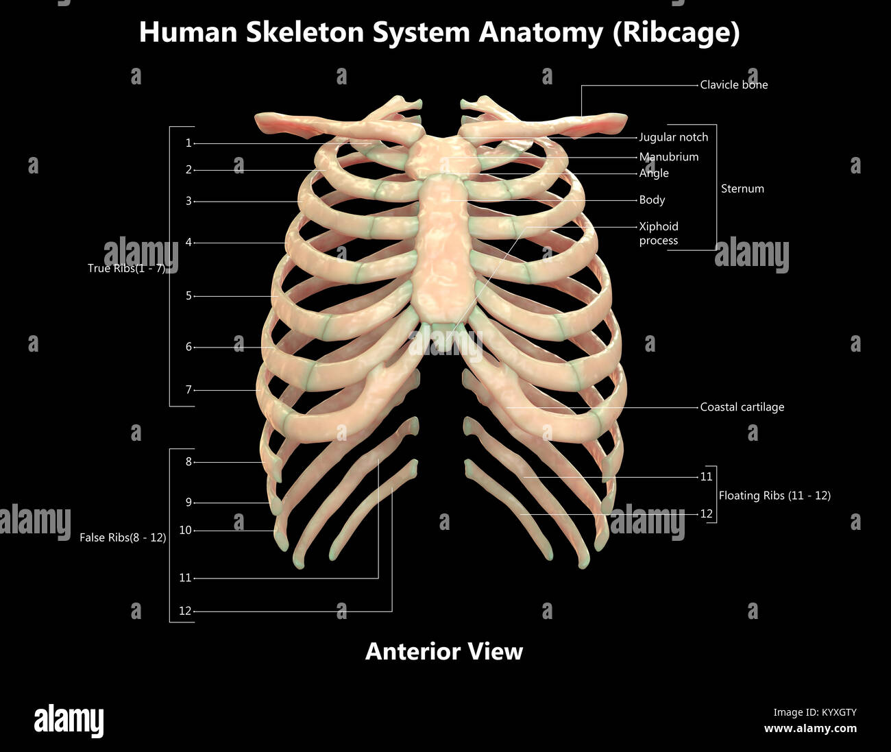
Human Skeleton System Rib Cage Label Design Anterior View ... from c8.alamy.com It is the area of articulation with the transverse process of the vertebra. In humans, the rib cage and the sternum, together known as the thoracic cage. The ribs are elastic arches of bone, which form a large part of the thoracic skeleton. In the anatomical position, the angles align with the medial border of the scapula. The part of the muscle is thought to depress the ribs. Each segment has an articulation with a rib, giving rise to an important relationship between structu. There are twelve pairs of ribs. Anatomy of the calf (posterior leg).
Ribs 3 to 9 are considered typical ribs.
Includes images, video, and free quiz. Ribs 3 to 9 are considered typical ribs. They also have a role in. It is split into ibrahim, af and darwish: .the anatomy of ribs as well as anatomy of rib cage in general. The anatomy of a typical thoracic vertebra is shown in figure 7.4a. Posterior interventricular branch of right coronary artery. The intercostal segment supplies the rib and adjacent muscle and other tissues. The anatomy of the ribs and their joints, forming the thoracic cage. The ribs partially enclose and protect the chest cavity, where many vital organs (including the heart and the lungs) are located. Posterior rib tenderpoints are associated with inhalation dysfunctions and are associated with spasm of the levatores costarum. This incision may be continued across the costal margin to open the abdominal cavity as in. The ribs stretches posteriorly from thoracic vertebrae to the anterior lateral edges of the sternum.
13 ribs anatomy and somatic dysfunctions anatomy of ribs. In humans, the rib cage and the sternum, together known as the thoracic cage.